General Details:
39 year old married female
Nipple-Areola Complex Sparing Mastectomy
General Details:
39 year old married female
Symptoms/ Signs:
Lump in Upper Inner Quadrant of Left Breast since 6 months
Swelling in Left hands since 6 months
Examination:
Definite single and palpable hard mass 2-5cms in size
No skin or nipple changes
No axillary lymph nodes
History:
No significant medical or gynaecologic history
Menstrual H/o – Normal
Family h/o bladder cancer in grandmother
Diagnostics:
Management:
Left Nipple-areola complex sparing mastectomy with subpectoral implant with Left sentinel node biopsy with Right Breast Mastopexy
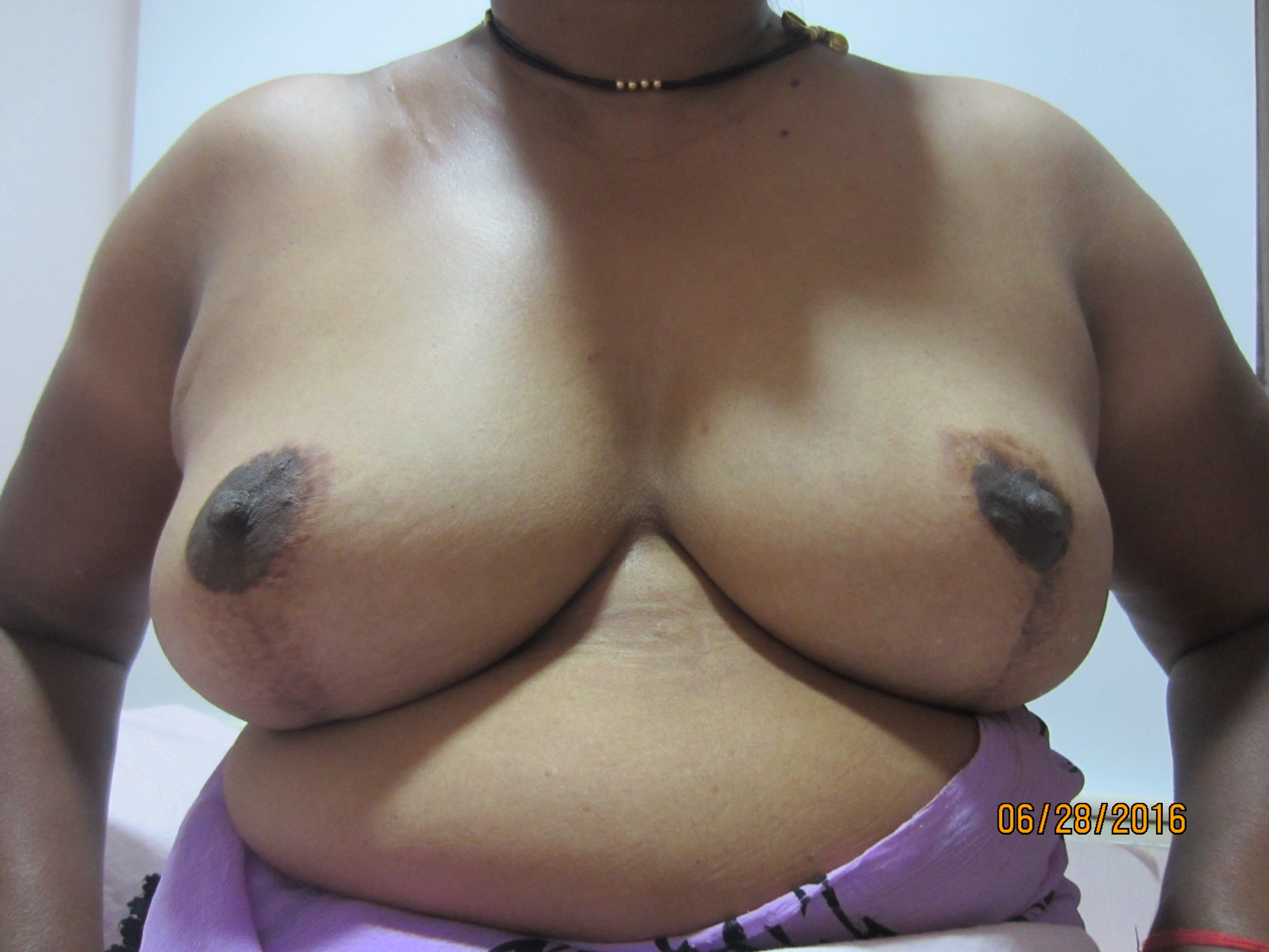
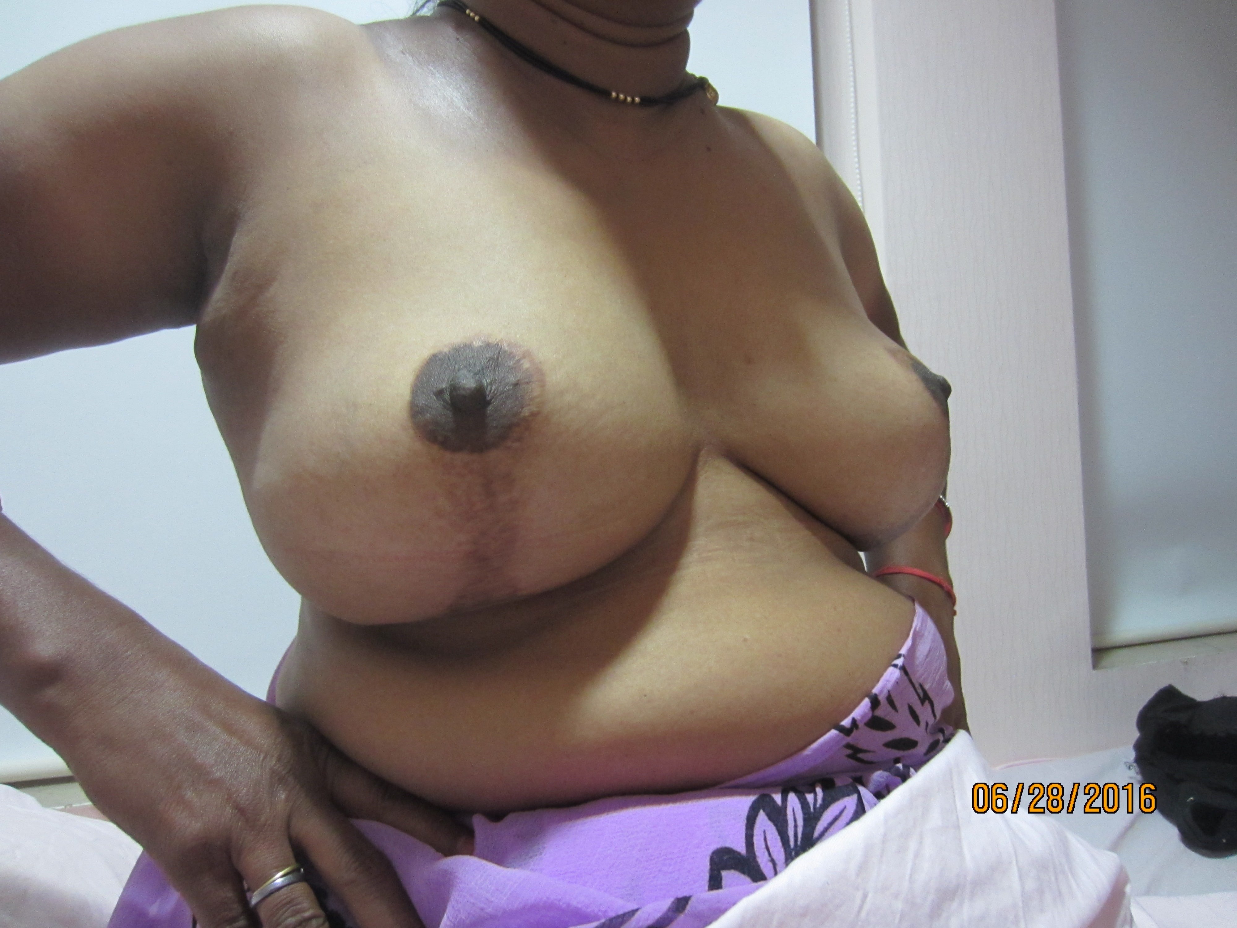
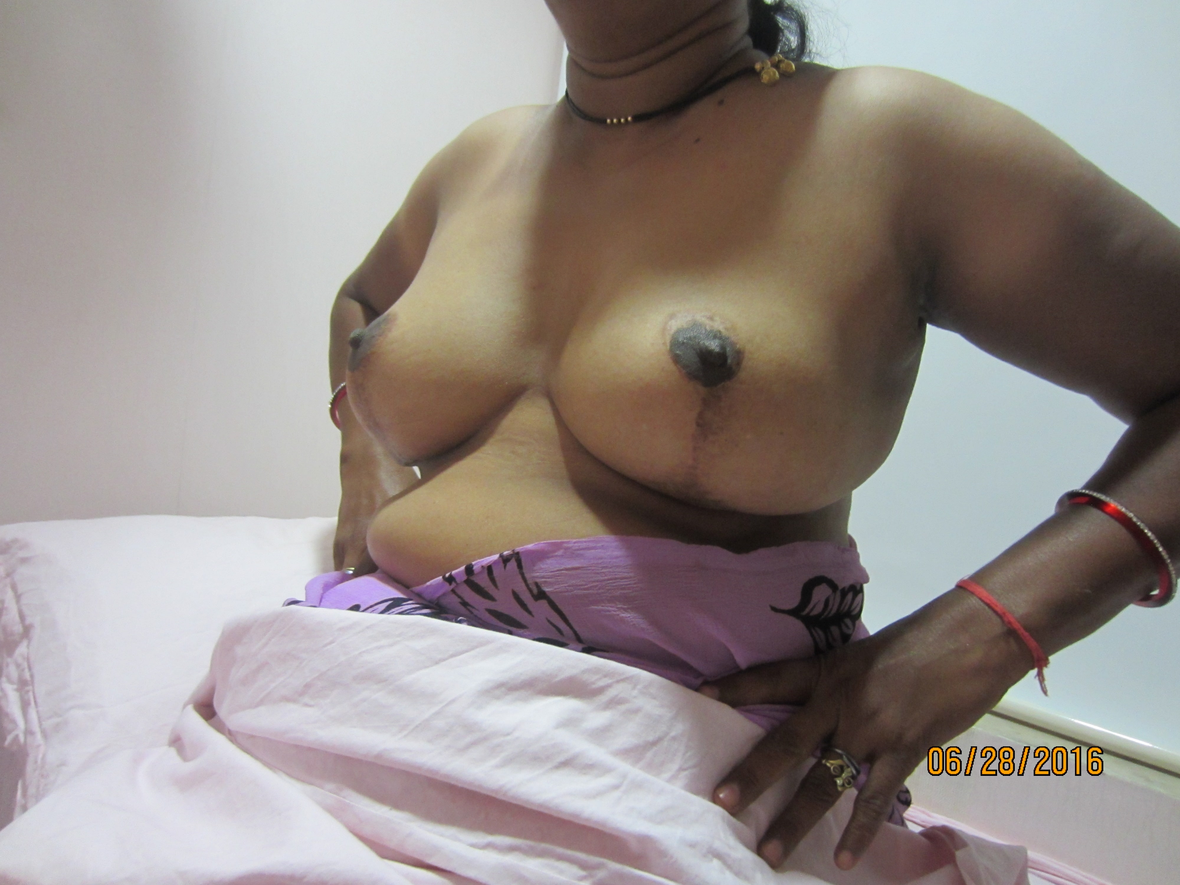
Discussion:
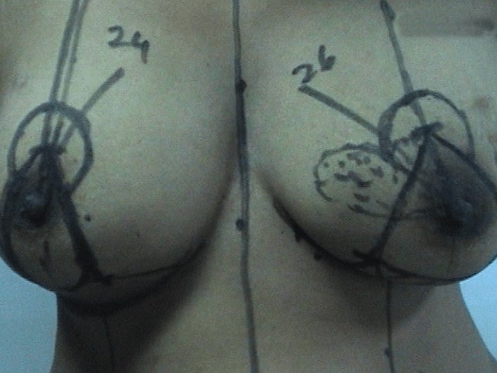
Nipple-Areola Complex (NAC) is at 26 cm from suprasternal notch on the Left Breast is and 24 cms
on the right side.
Tumour is at 9 o’clock position extending to 10 o’clock
Wise pattern marking is done
NAC is taken on superior pedicel
Inferior pedicle is converted into a sling
Mastectomy is commenced
Lower dermal sling is prepared from the lower segment
Flaps are raised medially and laterally and breast is removed
Breast after removal shows the patch of NAC
The nipple is cored out and NAC is dissected off
Mastectomy is completed and flaps are examined for uniformity
Flaps are dissected in the correct plane
Pectoralis muscle is raised, and the sling and Pectoralis Major are sutured together with Vicryl
The port is inserted and NAC is sutured back
Surgery conducted on the opposite breast:-
Opposite breast is reduced with a vertical scar technique using the superior pedicle carrying the
nipple-areola complex and the inferior pedicle is used for auto-augmentation
Inferior pedicle is fixed to the chest wall
The lateral and medial pillars are closed in the midline
T cut is given to remove the excess skin and to give a round shape
Surgical Histopathology Report:
Total excised mass: 500gm – 18cm x 18cm x 3cm
Original tumour size – 2cm x 2cm x 1.5cm
Margins: Free
Nodal involvement: 2 sentinel nodes negative for atypical or malignant cells
Tumour showed DCIS with central necrosis
On tissue blocks:-
ER/ PR - positive
HER 2 – negative
Adjuvant Hormonal Therapy:
Tab. Tamoxifen 20 mg once a day for 5 years